


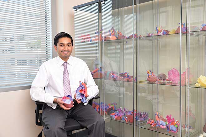
When a child is diagnosed with a serious health condition, every technological advantage counts. Highly advanced medical equipment, devices and procedures allow doctors to identify health problems sooner, develop the right treatments and save children’s lives. One of the most recent technologies to make its way into medical use is 3-D printing. Doctors are using it to create highly detailed, lifelike models of human organs and tissues to help plan surgeries, educate patients and their families, and train medical students and residents.
“Three-dimensional printing has been around for a while, but it’s only been used in cardiology for the past decade or so,” says Washington University physician Dr. Shafkat Anwar, cardiology director, cardiac MRI, at St. Louis Children’s and Washington University Heart Center. “These models are very helpful in showing patients what their surgery and treatment options are.” Doctors can bring the models to patient and family consultations to explain a disease, or show where an injury or congenital defect is and how it can be repaired, which helps boost patient confidence and reduce anxiety.
 “There are a couple of different models,” Anwar says. “We can make rigid ones out of resin or plaster. These can be colored and are great for teaching medical students about complex anatomy. We also can make models out of softer, more flexible materials that can be cut, opened up and manipulated. These are ideal in helping surgeons plan procedures before they enter the operating room.” No surgical case is cookie-cutter, so a clear and detailed model of the patient’s unique physiology can help surgeons create a plan A and plan B for each procedure, according to Anwar. “We can print a whole heart to look exactly as it would in the patient’s chest, including blood vessels,” he explains. “Having that information in advance helps us understand the disease process better, reduces time spent in the operating room, and hopefully leads to better outcomes for patients.”
Anwar says 3-D modeling is especially helpful in high-risk patients with life-threatening conditions. He gives the example of a patient needing heart surgery. “For procedures like this, we can create a whole chest model and take it into the operating room,” he notes. “Complex cardiac surgery is highrisk, even for senior surgeons. With a model, the surgeon can go through all steps of the procedure ahead of time. This is powerful technology that helps us deliver the best results for the patient.”
Anwar adds that the model-making process is pretty straightforward. Printing equipment is programmed with image data from a patient’s MRI or CT scan, and it uses that information to put down layer after layer of material until the model is complete.
To make modeling more widely available, St. Louis Children’s Hospital is building a new 3-D printing lab at the BJC Institute of Health at Washington University School of Medicine. Slated to open in early 2018, it is expected to include several printers and serve all 13 BJC hospitals. “We also anticipate being able to take model orders from other medical centers,” Anwar says. “The entire system is electronic, so we will be able to receive an MRI or CT scan of any patient instantly and ship out a finished model in a few days. We expect that 3-D modeling will become the standard of care, and we think it should be, for the good of patients.”
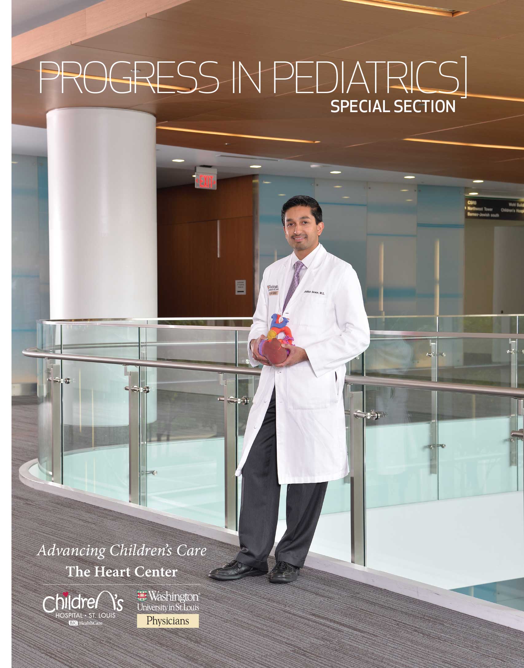
St. Louis Children’s Hospital, part of the BJC Healthcare System, provides comprehensive care for children from infancy to adolescence. Picture on the cover: Dr. Shafkat Anwar, Cardiology Director, Cardiac MRI, at St. Louis Children’s and Washington University Heart Center. For more information, call 314.454.6000 or visit stlouischildrens.org.
Cover design | Allie Bronsky
Cover photo | Bill Barrett
