


 For SLUCare neurosurgeon Dr. Philippe Mercier, it’s paramount to be able to differentiate a tumor from surrounding tissue during brain surgery, so advanced technology that can help him do that is extremely valuable. Mercier and his colleagues use a technique called intraoperative magnetic resonance imaging (iMRI), along with other technologies, to remove cancerous tissue without disturbing surrounding structures in the brain.
“Many people aren’t aware that tumor tissue often looks very similar to normal brain tissue,” Mercier says. “With iMRI, we can see subtle differences while a surgery is in progress so we can achieve what we call maximal safe resection, or the most effective removal of the tumor possible.” He explains that other factors like swelling or shifting of brain tissue also can make it difficult to assess a tumor’s location and size, and the technology helps to avert that issue as well.
Mercier says that in past decades, surgeons couldn’t always be certain that all of the affected tissue had been removed during an operation, so they might have to go back in later, which could introduce complications. With this advanced technology, however, they can be much more precise and help prevent later problems. “With some cancers such as those of the skin, it’s acceptable to take some surrounding tissue during a resection, but the same is not true of brain surgery,” Mercier explains. “We need to remove tumor tissue only, so we can be sure to preserve brain function.”
It’s not difficult to see why getting a resection right the first time is of such importance. “Imagine your child is in the hospital for surgery to remove a brain tumor,” explains Mercier, who practices at SSM Health Saint Louis University Hospital and SSM Health Cardinal Glennon Children’s Hospital. “No surgeon wants to close up the site, do post-procedure imaging and discover that some of the growth has been left behind. No one wants to have to tell that to a child’s parents. We can use iMRI in conjunction with other techniques like stereotactic guidance and microscopy with fluorescent filters to ensure the tumor has been successfully removed while the patient is still on the table.”
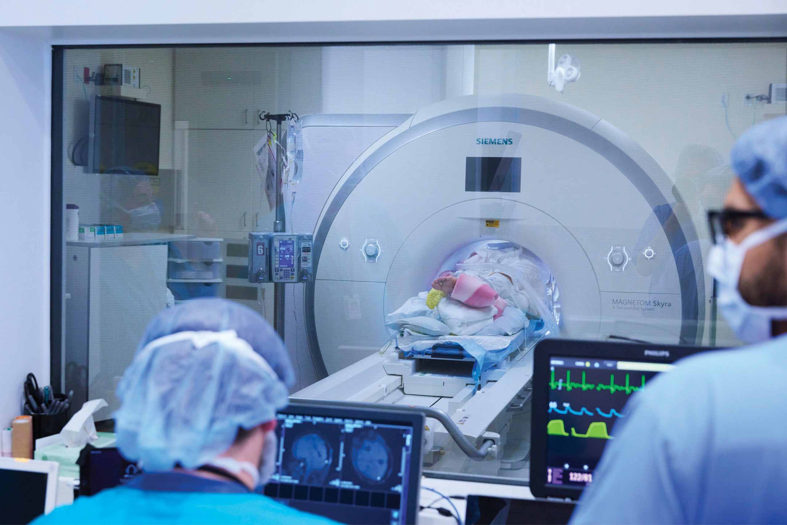
The iMRI scanner is situated between operating rooms so it can be used during more than one procedure at a time, Mercier says. “It’s state-of-the-art technology and a critical piece of equipment for the hospital,” he notes. “When we are finishing up with a surgery and want to make sure everything looks clear, we prep the patient, put him or her in the scanner and get imaging results quickly.”
Mercier says additional reasons to use iMRI are less invasiveness, an easier healing process and faster recovery times, and its use demonstrates SLUCare physicians’ dedication to the most effective ways to treat patients. “Neurosurgical oncology has changed drastically over the past 30 or 40 years,” he notes. “We are always adding and helping to develop state-of-the-art ways to achieve the best results for patients, and our surgeons have subspecialty training that allows us to target treatment to their individual needs. Technological advancements combined with personal expertise and skill make the difference for our patients.”
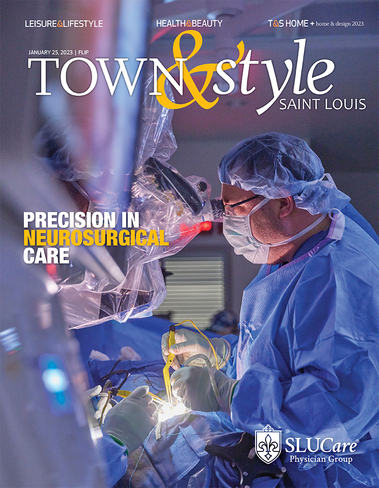
SLUCare physicians are committed to providing complete, individualized, state-of-the-art neurosurgical care for patients. Pictured on the cover: SLUCare neurosurgeon Dr. Philippe Mercier performs a procedure. For more information, visit slucare.com and search for ‘iMRI,’ or call 314.977.4440.
Cover design by Julie Streiler
Cover photo courtesy of SLUCare Physician Group
Pictured at the top: SLUCare neurosurgeons provide state-of-the-art care.
Photo courtesy of SLUCare Physician Group


